
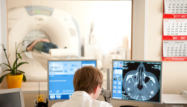
Patients with recurrent sinus infections often require more than a basic exam. Standard visual evaluations can assess the nasal passages, but they cannot examine the deeper sinus cavities. When facial pain, pressure, and congestion keep returning despite routine medical treatments, a deeper look is necessary to find out what CT scans reveal before endoscopic sinus surgery for recurrent sinus infections.
A sinus CT scan provides detailed imaging of the sinus anatomy and underlying disease. This advanced sinus imaging uses computerized X-ray technology to build clear cross-sectional images of the bone, soft tissue, and air spaces. This detailed imaging helps determine whether structural problems are contributing to chronic symptoms.
Without this detailed map, diagnosing the true source of your discomfort is mostly guesswork. These detailed scans play a major role in planning safe, targeted endoscopic sinus surgery, giving your ENT specialist a roadmap to clear your airways. If you are seeking CT scan sinus surgery in Los Angeles, understanding these findings is an important first step.
Why CT Scans Are Important Before Sinus Surgery
The sinus system is a complex network of air-filled pockets located behind your forehead, cheeks, and eyes. A standard physical exam cannot see into these deep spaces. This explains why CT scans are important before sinus surgery, as they provide a clear view of every nook and cranny, mapping your entire nasal passages.
Many people suffer from physical blockages that they were born with or developed over time. These include malformed bone structures or blocked drainage pathways that cannot be detected with a standard nasal speculum. The scan brings these hidden issues to light.
Swollen tissues can block the normal flow of mucus, leading to recurrent sinus infections. Imaging shows exactly which sinus linings are thick, inflamed, or fluid-filled, differentiating between healthy air spaces and diseased tissue.
Surgery is not always the answer for every sinus issue. A clear scan lets your physician see whether your symptoms are caused by a structural defect that requires mechanical correction or if your issues are purely medical and better managed with medication.
Every person has a slightly different internal nasal structure. Operating without a scan would be like driving through an unfamiliar city without a map. These images guide the surgeon’s path, protecting delicate tissues and focusing only on the areas that need repair.
Also Read: Opening Blocked Nasal Passages: 4 Invaluable Benefits of Endoscopic Sinus Surgery
What CT Scans Can Reveal in Patients With Recurrent Sinus Infections
Blocked Sinus Drainage Pathways
The sinuses depend on tiny, natural openings to drain mucus and stay healthy. If these channels become blocked, fluid traps bacteria and creates an ideal environment for infections. A scan shows areas where mucus cannot properly drain, highlighting the exact locations where blockages occur. By identifying a chronic sinus blockage diagnosis with CT imaging, the doctor can pinpoint the exact sites that require surgical opening.
Chronic Sinus Inflammation
When infections keep coming back, the delicate tissue lining the sinuses undergoes changes. This is a primary component of a chronic sinusitis diagnosis with CT imaging in Los Angeles practices. The scan detects thickened sinus lining and fluid buildup that marks ongoing disease. This visualization helps distinguish chronic sinusitis from a temporary infection, showing whether your tissues are permanently altered or simply dealing with a brief cold.
Deviated Septum
The nasal septum is the wall of bone and cartilage that divides your nasal cavity in half. When this wall leans significantly to one side, it restricts airflow and blocks mucus clearance. The imaging reveals narrowing of nasal passages caused by this lean. It clearly shows how septal deviation affects airflow and drainage, allowing the surgeon to see how correcting the bone will balance your breathing.
Nasal Polyps
Nasal polyps are noncancerous, teardrop-shaped growths that develop on the lining of your nasal passages or sinuses. They often result from chronic swelling due to asthma, recurring infections, or allergies. When analyzing sinus CT scan findings before endoscopic sinus surgery, a doctor looks closely for these tissue masses. The scan identifies inflammatory growths blocking the sinuses, showing where these polyps reside and revealing if a polyp is small or if it fills an entire sinus cavity.
Narrow Sinus Anatomy
Sometimes, the root cause of chronic sinus trouble is simply genetics. Some individuals are born with naturally tight sinus openings that clog at the slightest hint of inflammation. The scan detects naturally tight sinus openings easily. It reveals anatomical variations affecting drainage, allowing your doctor to see if your body’s natural architecture is predisposed to blockage.
Hidden Structural Abnormalities
Nasal trauma, old injuries, or unique anatomical variations can alter the internal structures of your face. A detailed evaluation of recurrent sinus infections and CT scan evaluation records can uncover these issues. The scan identifies bone irregularities or prior scar tissue that might be narrowing your air passages. It detects issues not visible during a routine exam, finding deep bone spurs or unusual sinus cells that would otherwise remain invisible.
How CT Imaging Helps Plan Endoscopic Sinus Surgery
Maps the patient’s unique sinus anatomy
No two human skulls are identical inside. A scan acts as a personalized GPS, mapping your specific sinus paths. The surgeon reviews these images prior to your procedure to understand your exact nasal landscape, identifying where your sinuses are wide, narrow, or asymmetrical.
Helps surgeons avoid critical structures
The sinus cavities sit directly adjacent to highly delicate zones, including the eye sockets, optic nerves, and the base of the skull. Imaging helps surgeons avoid these sensitive structures by clearly marking the boundaries between your sinuses and these vital areas, protecting your vision and neurological health.
Guides minimally invasive surgical techniques
Endoscopic sinus surgery utilizes small, lighted tubes inserted through the nostrils, requiring no external incisions. The detailed map provided by the scan guides these minimally invasive surgical techniques. Your surgeon can navigate directly to the trouble spots without disturbing healthy tissues nearby.
Improves surgical precision and safety
Operating with a highly detailed map improves overall surgical accuracy. Your physician can clear blockages, remove polyps, and widen narrow passages with millimeter-level accuracy, lowering the risk of accidental tissue damage and making the entire procedure much safer.
Helps determine the extent of surgery needed
Some patients only need one specific sinus cavity opened, while others require treatment across their entire sinus network. Looking at the scan helps determine the extent of surgery needed, preventing undertreatment of hidden issues while avoiding unnecessary work on healthy areas.
Also Read: The Cure for Chronic Sinusitis
Why Physical Exams Alone Are Not Enough
A routine office evaluation typically involves a doctor looking up your nose with a small light or a flexible endoscope. While this is a helpful starting point, some sinus problems cannot be seen during a routine office exam. Deep sinus passages sit inches behind the face, far out of reach of direct sight lines.
To get a true picture of your health, doctors rely on specific clues regarding what ENT specialists look for on sinus CT scans, such as deep tissue thickening and blocked passageways. Symptoms alone do not always reveal the underlying cause. A patient with severe facial pressure might have a massive polyp, or they might simply have a severe allergic attack. A scan provides objective imaging of chronic disease, taking away the guesswork. This imaging helps pave the way for an accurate diagnosis and treatment plan, building a strategy based on clear visual evidence rather than symptoms alone.
What to Expect During a Sinus CT Scan
If you have never had a scan of your facial bones, there is no need to feel anxious. The process is a quick, noninvasive imaging process that requires very little effort from the patient. You will simply lie on a motorized table that slides your head into a large, doughnut-shaped machine.
The scan typically takes just a few minutes, during which you will need to remain completely still. No surgical preparation is required, so you do not need to fast or stop taking your daily medications beforehand. The advanced machine produces highly detailed images of the sinus cavities, which are sent directly to a computer for your ENT specialist to review.
When CT Scans Are Typically Recommended
A sinus scan is not the first step for a standard cold, but it becomes necessary when sinus issues become unmanageable. Doctors typically recommend this step when a patient experiences:
- Recurrent sinus infections that return multiple times a year
- Chronic sinusitis symptoms lasting 12+ weeks without relief
- A poor response to standard medical therapies like antibiotics, steroid sprays, or allergy pills
- A suspected structural blockage, such as a severely deviated septum or visible polyps
- An upcoming evaluation before considering endoscopic sinus surgery
How CT Findings Influence Treatment Decisions
The images from your scan determine your next medical steps. First, the images determine whether surgery is necessary or if your sinuses can still be managed with modified medical treatments. If surgery is selected, the scan helps decide which structures need correction, ensuring the surgeon focuses on the exact areas causing your blockages.
This objective data supports a personalized treatment plan built around your specific anatomy. By showing exactly where the problems lie, this targeted approach may prevent unnecessary procedures on healthy sinus cavities, keeping your medical journey as straightforward as possible.
Benefits of Image-Guided Surgical Planning
Using a detailed scan prior to your operation changes the entire patient experience. The primary benefit is a more accurate treatment, as your surgeon can target the exact roots of your physical blockages. This leads directly to improved sinus drainage, allowing your body to clear mucus naturally as it was designed to do.
Over time, this results in better long-term symptom relief, breaking the cycle of constant pressure, pain, and antibiotics. Additionally, having a clear anatomical map reduces the risk of complications during the procedure, leading to excellent surgical outcomes and a smoother, faster recovery at home.
Also Read: Is Sinus Surgery Your Solution? Understanding the Path to Relief
Conclusion
CT scans are a fundamental step before endoscopic sinus surgery. They remove the guesswork from your sinus care, turning hidden physical blockages into clear, viewable maps. This imaging reveals structural problems and chronic inflammation that may be causing recurrent sinus infections, showing your doctor exactly why medications have failed to give you lasting relief.
A detailed evaluation allows for safer, more precise treatment planning, keeping your safety and comfort at the forefront of the procedure. If you are tired of living with constant facial pressure, congestion, and frequent infections, it is time to seek answers. Schedule a consultation with Dr. Mani H. Zadeh, MD, F.A.C.S., for an expert sinus evaluation and personalized care.